Radiography is one of the oldest, most approved and used, diagnostic imaging techniques in veterinary medicine. The Bargteheide Equine Hospital possesses two stationary digital x-ray generators as well as a portable machine for theatre use or horses being high grade lame so that they cannot walk into x-ray room.
The use of radiography encompasses nearly every field in veterinary medicine. Lameness examinations require radiographs of different parts of the limb, a standard prepurchase examination contains radiographs beside the clinical investigation and in patients with misbehavior when being ridden we generate x-rays of the back and neck. With our x-ray generator we are also able to obtain good quality radiographs of the hip joint in the standing horse.
In patients with dental or skull diseases radiography is helpful to get more information about dental roots as well as frontal and paranasal sinuses and the nasal cavity. Thoracic radiography is the diagnostic method of choice for evaluating pulmonary parenchymal disorders.
Contrast radiographs are generated in ataxic horses (myelography) with suspicion of cervical vertebral stenotic myelopathy or in cases of a questionable soft tissue injury of a joint (artrography). But radiography can also be a helpful tool to control the exact needle position for medical injections or to document treatment progresses.
To obtain best quality radiographs and stress your horse as less as possible nearly every horse will be sedated in x-ray room. Every radiograph will be archived for 10 years and if you wish you can receive your horses x-rays on a CD (with little amount of costs).
-
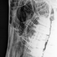
- head: lateral view
-
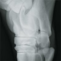
- hock joint
-

- Navicular bone oxspring
-
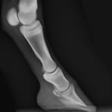
- foot and fetlock lateral
-
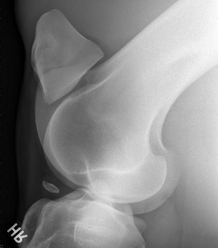
- stifle joint


