Magnetic resonance imaging is one of the newest diagnostic techniques in equine medicine. With the advent of diagnostic quality standing equipment this technology has increasing utilisation in the field of equine orthopaedics.
MRI does not use ionizing radiation to produce an image. Three major components – hydrogen atoms in the tissues, a strong external magnet and an intermittent radio wave – are required to generate images based on differing magnetic properties of tissues.
Its main area of use has been in the hoof capsule where ultrasonographic examination is limited. However the additional information it can yield can also be beneficial in scanning the pastern, fetlock, suspensory and carpal and tarsal regions. MRI is not useful as a screening procedure and is best employed in assessing as small an area as possible
- MRI hoof
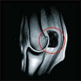
- sclerosis of the proximal semoid bone fetlock sagittal slice
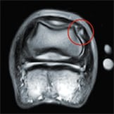
- collateral ligament lesion horizontal slice
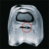
- core lesion DDFT transverse slice
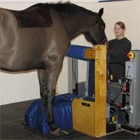
- MRI examination


